
Step-by-step guide to conducting fiber microtome test
Here is a step-by-step guide to conducting a fiber microtome test:
1. Sample Preparation: Prepare the fiber sample according to the test requirements. This may involve selecting and separating individual fibers, aligning them for cutting, and embedding them in a suitable medium.
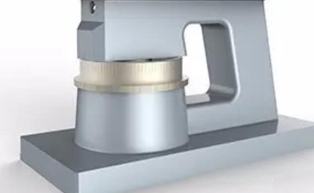
2. Mounting the Sample: Mount the prepared fiber sample onto the sample holder of the microtome using adhesive or clamps. The sample should be securely fixed to prevent movement during cutting.
3. Adjusting the Blade: Adjust the blade to the desired thickness of the fiber section to be cut. This can be done by adjusting the blade holder or by using a micrometer.
4. Cutting the Fiber: Move the sample holder into position under the blade and start the cutting process. The blade should be moved through the fiber sample at a controlled speed to ensure consistent thickness of the sections.
5. Collecting the Sections: Collect the sections of fiber as they are cut, and place them onto glass slides for microscopic analysis. The sections should be handled carefully to prevent damage or distortion.
6. Staining and Mounting the Sections: Stain the sections of fiber using appropriate staining techniques to enhance contrast and visibility under the microscope. Mount the sections onto glass slides using a suitable mounting medium.
7. Microscopic Analysis: Examine the mounted sections of fiber under a microscope to study their morphology, structure, and composition. Measure and record any relevant parameters or characteristics.
8. Reporting: Report the test results in a clear and concise manner, including the test conditions, measurement data, and analysis. The report should also include any observations or deviations from the test method specification.
In summary, conducting a fiber microtome test involves preparing the fiber sample, mounting it onto the microtome, adjusting the blade, cutting the fiber, collecting the sections, staining and mounting the sections onto glass slides, microscopic analysis, and reporting the results. By following these steps, it is possible to obtain reliable and accurate data on the morphology, structure, and composition of fibers, leading to a better understanding of their properties and characteristics.